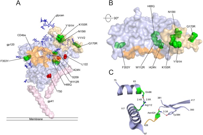
Fig 5. Mapping of mutations on gp140 structure.
(A) Surface view of the structure of a BG505 SOSIP.664 gp140 monomer. The gp120 unit is shown in light blue and the gp41 unit in light pink. Glycans are shown as blue sticks, the CD4bs is orange and the V1/2 loop domain golden. Mutated residues found in clone 3.3.1 only are shown in red and residues found in clones 4.3.B01 and 4.3.D01 are shown in green. (B) The molecule was rotated 90° around the x-axis and residues found in 4.3.B01 and 4.3.D01 are shown in green. (C) Cartoon view of mutated residues found within gp120, putative hydrogen bonds are denoted as dashed lines.

