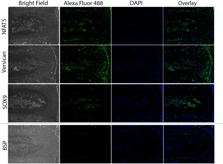
Fig 2. Immunohistochemical staining validating the localization of proteins in the murine intervertebral disc.
Representative images demonstrating the immunolocalization of NFAT5, versican, Sox9, and BSP within the murine IVD. Each protein demonstrates a distinct pattern of localization within specific compartments of the disc. NFAT5 is localized to the NP and CEP, versican is present throughout the disc with high levels detected in the outer annulus, Sox9 is present in NP and CEP and BSP is only detected in the CEP. For each protein-specific antibody, sections corresponding to 3 IVDs were assessed for each animal; n = 3 mice. Scale bar = 100 μm.

