
Figure 2.
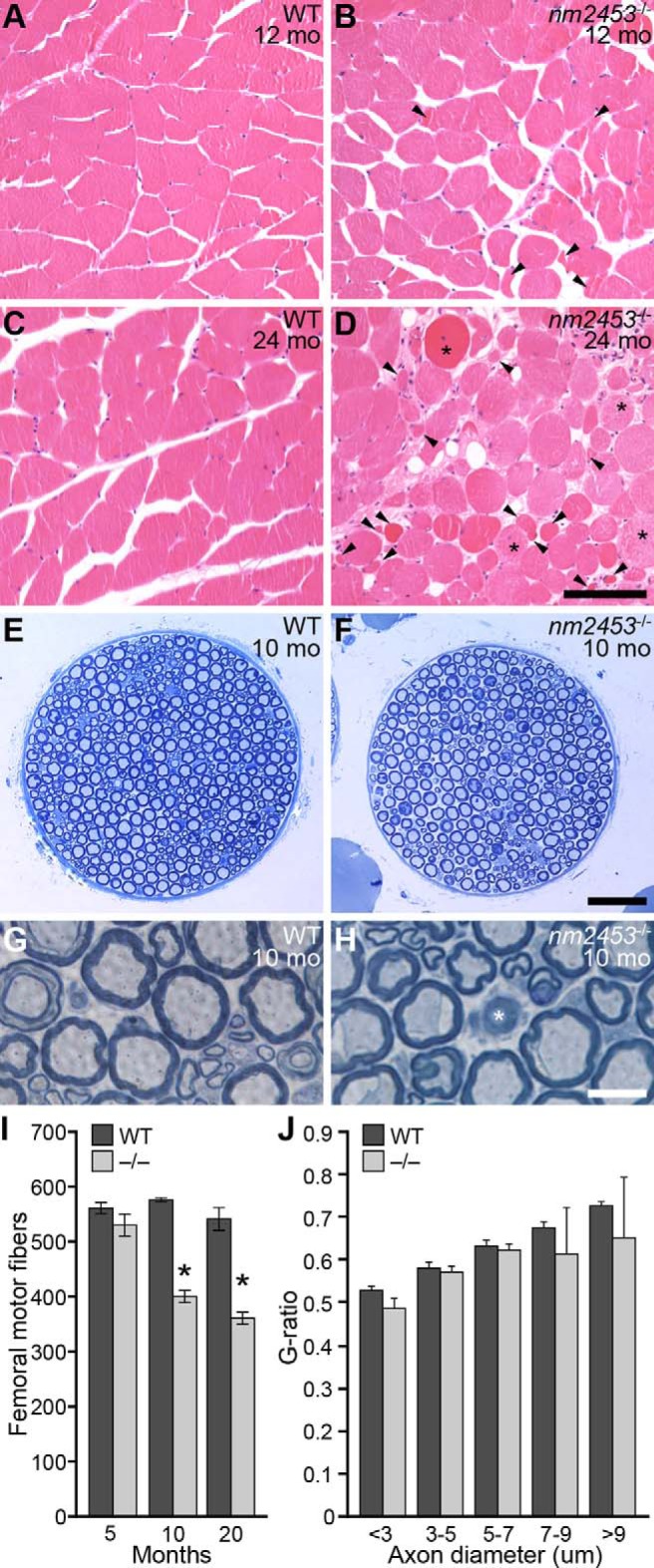
Muscle atrophy and peripheral neuropathy in nm2453 mutant mice. A–D, Hematoxylin- and eosin-stained cross sections of gastrocnemius muscles from wild-type (WT) and nm2453−/− mice. Arrowheads and asterisks indicate atrophied muscle fibers and fibers with central nuclei, respectively (D). E–H, Toluidine blue-stained cross sections of femoral nerve motor branches from 10-month-old WT and mutant mice. Axon degeneration (asterisk), but no typical signs of demyelination, was observed as shown in higher-magnification images (G, H). I, Quantitation of axon numbers in the motor branch of the WT and mutant femoral nerve from 5-, 10-, and 20-month-old mice. J, The g-ratio of axons in the motor branch of the femoral nerve from 12-month-old WT and nm2453−/− mice. No significant changes were observed between WT and mutant axons. Results in I and J are presented as mean ± SEM (n = 3). *p < 0.01 (statistically significant differences between similarly aged mutant and WT mice and aged and 2-month-old mutant mice). Scale bars: D, 100 μm; F, 50 μm; H, 10 μm.