Figure 5.
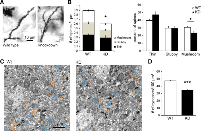
Chmp2b is involved in the maintenance of excitatory synapses in vivo. A, Brain sections from 5-week-old Chmp2b knockdown mice (KD) or sibling controls (Wild type) were stained by the Golgi impregnation technique. Stained dendritic segments were visualized by bright-field microscopy of the stratum radiatum. Shown are representative images of dendritic shafts and spines in both genotypes. B, Spines were classified into thin, stubby, or mushroom types, and their density per unit dendritic length was measured for each type (n = 4 animals, 36 dendrites per genotype). Left, Histogram showing the mean density of each spine type in KD and WT mice. The total spine density was significantly lower in KD mice than in controls (*p = 0.039, two-sided t test). Right, Histogram showing the mean proportion of the three spine types. The percentage of mushroom spines was significantly reduced in the KD mice (*p = 0.02). C, Representative electron micrographs of the CA1 stratum radiatum from Wt and KD siblings. Presynaptic profiles are highlighted in blue, and postsynaptic spines (s) are in brown. D, Quantification of synapse density in the electron micrographs. WT: n = 3 animals, 851 axo-spinous synapses from 30 micrographs; KD: n = 3 animals, 627 axo-spinous synapses from 30 micrographs. The density of postsynaptic spines per unit surface was significantly lower in the KD (***p = 0.0008).

