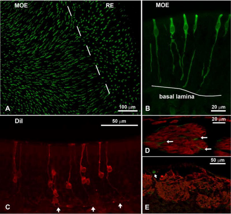
Fig. 2.

Confocal micrographs of IP3R3-tauGFP mice showing the olfactory epithelium (A-C), fila olfactoria (D), and the olfactory bulb (E). A) Whole-mount en face view of IP3R3 MV cells in an area where the main olfactory epithelium (MOE) and the respiratory epithelium (RE) meet. The IP3R3 MV cells are longer and more abundant in the MOE than in the RE. B) Higher magnification of the IP3R3 MV cells in a cross-section through the MOE. The cells stretch the whole height of the epithelium but the basal processes do not pass the basal lamina. The cell body lies in the upper third of the epithelium. C Cells retrogradely labeled with DiI from the olfactory bulb show the typical morphology of olfactory neurons (OSNs): a long thin dendrite; a cell body located in the lower portion of the epithelium; a thin axon projecting to and passing the basal lamina (arrows). D Antibodies against OMP show fila olfactoria beneath the MOE (red). Within the bundles, few IP3R3-positive fibers (green) of unknown origin are visible that do not double-label with OMP (arrows). E Section of the olfactory bulb where glomeruli are labeled with OMP. Note that the fibers of the glomeruli do not express IP3R3. However, small blood vessels show GFP labeling (arrow).
