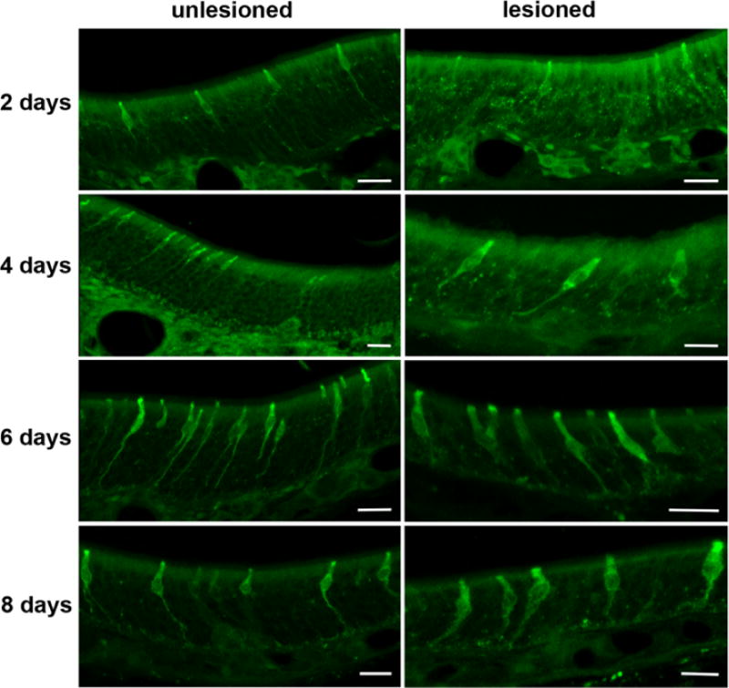
Fig. 4.

Unilateral bulbectomy revealed that the IP3R3 MV cells do not degenerate. Representative confocal z-stack images of the MOE 2 – 8 days post-bulbectomy are shown. The lesioned and the unlesioned control side contained a comparable amount of IP3R3 MV cells during the entire experiment as seen in these micrographs for days 2 to 8 after surgery. All scale bars = 20 μm.
