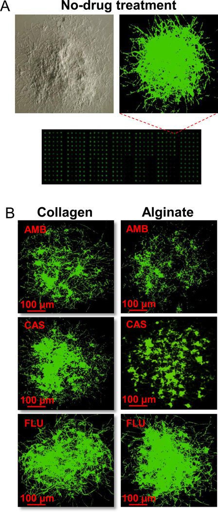
Figure 1.
A microarray scanner image of Candida albicans nano-biofilms. (A) Light and fluorescence microscopic images of a mature C.albicans nano-biofilm stained with FUN1. (B) A panel of drug-treated nano-biofilms encapsulated in collagen and alginate hydrogels. The nano-biofilms were treated for 24 h with amphotericin B (AMB), caspofungin (CAS) or fluconazole (FLU) at 1 µg/ml, 3 µg/ml and 1024 µg/ml, respectively, stained with FUN1, and imaged using a microarray scanner. Filamentous hyphae can be seen attesting the presence of true biofilms.

