Figure 8.

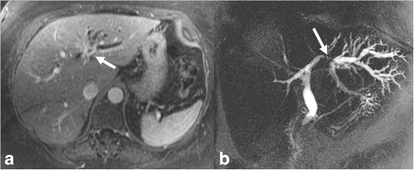
Infiltrating pCCA. Axial post contrast enhanced T1-weighted MR image (a) and MRCP (b) images demonstrating an enhancing stricture involving the left hepatic duct (arrow) with upstream dilatation of the left hepatic ducts.

Infiltrating pCCA. Axial post contrast enhanced T1-weighted MR image (a) and MRCP (b) images demonstrating an enhancing stricture involving the left hepatic duct (arrow) with upstream dilatation of the left hepatic ducts.