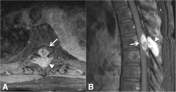
Figure 1.

A 61-year-old woman with breast cancer metastatic to the spine leading to spinal cord compression syndrome. Axial (A) and sagittal (B) post-contrast T1-weighted MR images of a patient with metastatic breast cancer showing a bone lesion in the posterior elements of T6 (arrowhead), with high T2 signal intensity and intense contrast enhancement, impressing upon the spinal canal and dislocating the cord anterolaterally (arrow).
