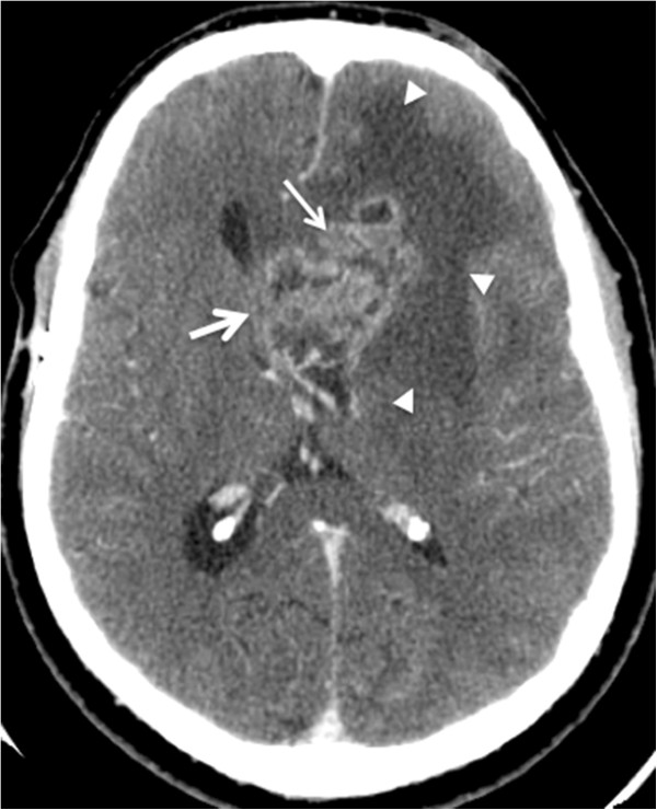
Figure 2.

A 37-year-old man with intracranial tumor with mass effect. Axial post-intravenous contrast images. Computed tomographic images of a patient with glioblastoma multiforme in the left frontal lobe. The lesion with irregular contours and heterogeneous enhancement (thin arrow) has mass effect characterized by hypoattenuation (edema) of the adjacent white matter (arrowheads), compression of the lateral ventricle, and contralateral deviation of the medial line structures, with signs of subfalcine herniation (thick arrow).
