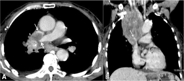
Figure 3.

A 55-year-old man with superior vena cava syndrome. Computed tomographic images of a patient with metastatic laryngeal cancer showing an infiltrative mediastinal mass causing compression of the right pulmonary artery (thick arrow) and superior vena cava (thin arrow). (A) Post-contrast axial slice. (B) Post-contrast coronal reconstruction.
