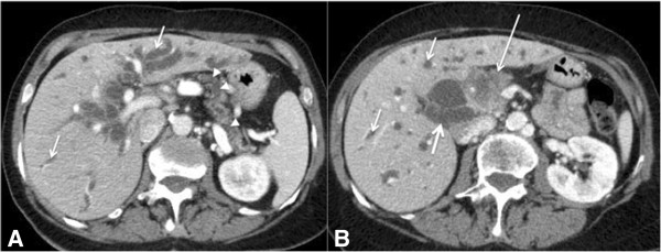
Figure 9.

A 58-year-old man with biliary obstruction caused by adenocarcinoma of the pancreas. Axial (A) and (B) CT images of the abdomen obtained after intravenous contrast administration demonstrating dilatation of the intra- (thin arrows) and extrahepatic bile ducts (thick arrow), a pancreatic head mass (adenocarcinoma) with irregular contours and poorly defined borders (long arrow), and dilatation of the pancreatic duct (arrowheads).
