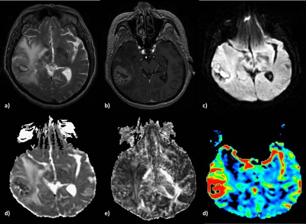
Figure 2.

Anaplastic Astrocytoma in a 71-year-old man. a) T2-weighted image shows increased signal intensity with peritumoral edema, b) heterogeneous contrast enhancement on a post-contrast 3D-SPGR image and c) restricted diffusion in the solid portion of the tumor. The lesion is hypointense on the ADC map (d), presents low FA (e) and increased perfusion on the rCBV map (f).
