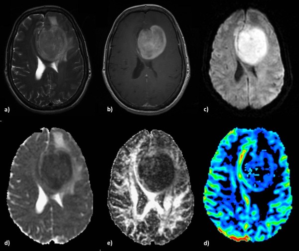
Figure 6.

Primary Cerebral Lymphoma in a 59-year-old-woman. a) High signal intensity with peritumoral edema on a T2-weighted image, b) intense contrast-enhancement on a T1-weighted post contrast image and c) hyperintensity on a DW image. Decreased intratumoral ADC (d) and FA (e). The rCBV map shows moderate perfusion within the lesion (f).
