
Fig. 4.
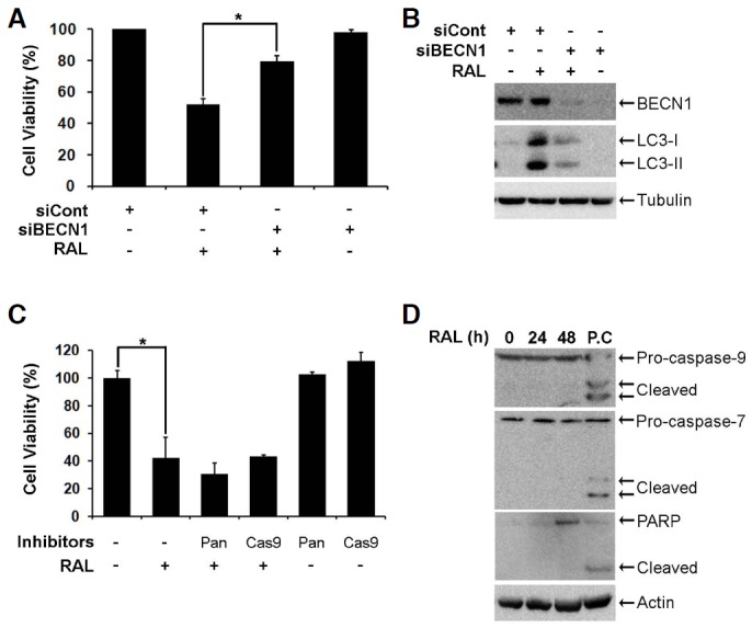
Raloxifene induces autophagy-dependent cell death. (A) MCF-7 cells were transfected with 0.17 μM non-targeting control siRNA (siCont) or BECN1 siRNA (siBECN1) for 48 h. Bars denote cell viability of cells treated with 10 μM raloxifene for 48 h, and cell viability was assessed using the MTS assay (mean ± SD; n = 3). *p < 0.05 according to one-way ANOVA. (B) MCF-7 cells were transfected with 0.17 μM siCont or siBECN1 for 48 h. BECN1 and LC3 were analyzed using Western blot analysis. (C) MCF-7 cells were pretreated with 20 μM caspase inhibitors for 2 h and then exposed to 10 μM raloxifene for 48 h. Cell viability was measured using the MTS assay (mean ± SD; n = 3). *p < 0.05 according to one-way ANOVA. (D) MCF-7 cells were treated with 10 μM raloxifene for the indicated times. Caspase-7, -9, and PARP were analyzed using Western blotting. The lysate of the HCT116 cells treated with 10 μM PXD101 for 24 h was used as a positive control (P.C) to assess the cleavage of caspase-7, -9, and PARP.