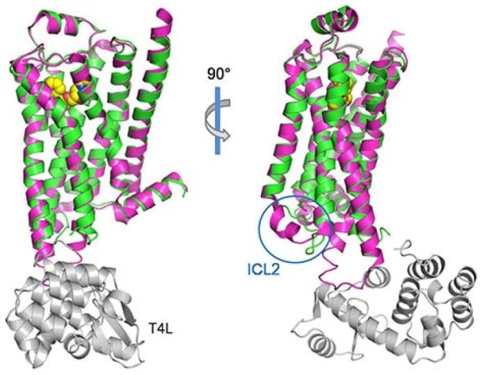
Fig. 5.

The superposition of β1AR from the antagonist bound inactive structure into the carazolol bound β2AR with T4L fusion. The structure of β1AR (pdb ID: 2YCW) is shown in magenta and the β2AR structure is colored as in Fig. 1A. The two structures are very similar except for the intracellular loop 2 (ICL2), circled in blue. Unlike in β2AR, ICL2 forms an α helix in β1AR.
