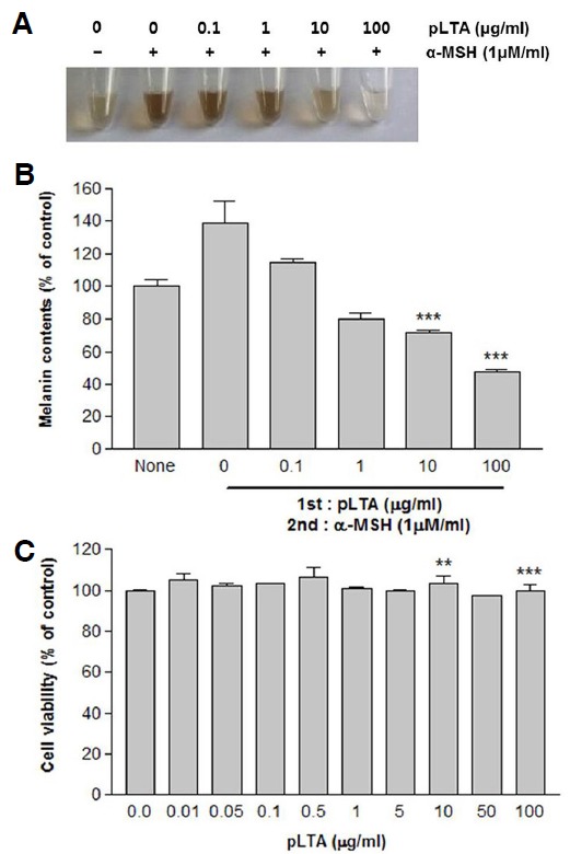
Fig. 1.

Effect of pLTA on melanin synthesis in B16F10 cells. The 1 × 10⁵ B16F10 cells (n = 3) were incubated with the indicated concentrations of pLTA for 72 h. Cells were dissolved in 1N NaOH and photographed (A). Alternatively, melanin content was determined as described in the “Materials and Methods” (B). Cell viability was determined by WST-1 assays (C). Results are the means of three independent experiments (mean ± S.D). ***P < 0.001 versus α-MSH treated cells.
