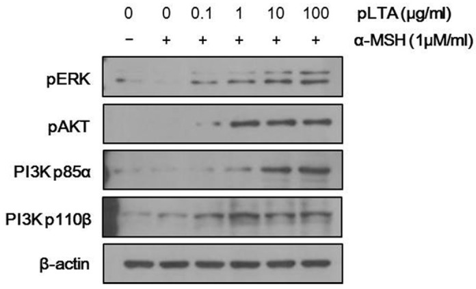
Fig. 4.

Activation of ERK and PI3K/AKT signaling by pLTA stimulation. The 1 × 10⁵ B16F10 cells (n = 3) were incubated at the indicated concentrations of pLTA for 24 h, and then stimulated with α-MSH for 2 h. Levels of p-ERK and p-PI3K/AKT were measured by Western blot analysis. To verify the amount of loaded protein, the blots were also probed with anti-β-actin.
