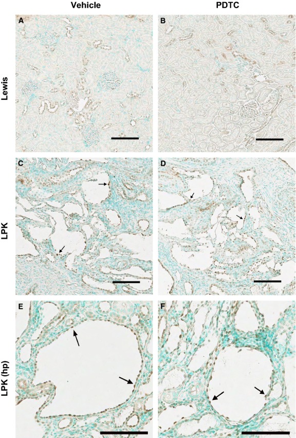
Figure 6.

Effect of PDTC on cortical P‐p105 immunolocalization. Shown are kidney cortices from Lewis rats treated with vehicle (A) and PDTC(40 × 2) (B), and LPK rats treated with vehicle (C, E) and PDTC(40 × 2) (D, F). Lewis cortices displayed P‐p105 positivity in distal tubule nuclei. In LPK kidney cortices, P‐p105 stained strongly in cystic epithelial cell (CEC) cytoplasm and nuclei (arrows). Scale bar = 100 μm.
