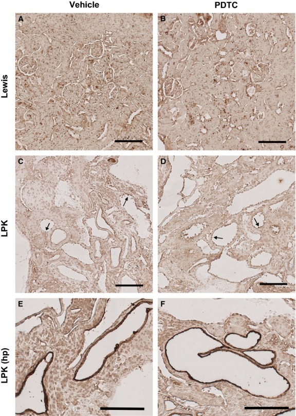
Figure 8.

Effect of PDTC on cortical p50 immunolocalization. The renal cortex is shown from a Lewis rat treated with vehicle (A) and PDTC(40 × 2) (B), and Lewis polycystic kidney (LPK) rats treated with vehicle (C, E) and PDTC(40 × 2) (D, F). Lewis cortices displayed p50 positivity in distal tubule nuclei. In LPK cortices, moderately positive staining was observed in the cytoplasm and nuclei of cystic epithelial cells (CECs) (arrows), and in distal tubule nuclei. There was occasional strongly positive staining in the CECs of focal cysts (E and F). Scale bar = 100 μm.
