Figure 1.
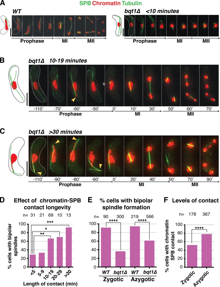
Rescue of bqt1Δ spindle defect by prophase chromatin–SPB contacts. (A–C) Frames from films of meiocytes carrying Hht1-mRFP (histone H3 tagged at one of the two endogenous hht1+ loci; Chromatin), Sid4-GFP (endogenously tagged; SPB), and ectopically expressed GFP-Atb2 (nmt1 promoter controlled; Tubulin). Numbering indicates meiotic progression in minutes; t = 0 is just before spindle formation. Bars, 5 µm. (A) A bqt1Δ meiocyte with <10-min contact displays a monopolar MI spindle and unstable MII spindles. (B and C) bqt1Δ cells with 10–19-min and >30-min chromatin–SPB contact (yellow arrowheads) show proper spindles at MI and MII. (D) Quantitation of effect of chromatin–SPB contact duration on bipolar MI spindle formation. n is the total number of cells scored in eight independent experiments; data were subject to Fisher’s exact test: ***, 0.0001 < P < 0.001; **, 0.001 < P < 0.01; *, 0.01 < P < 0.05. All cells scored for A–D of this figure are more extensively analyzed in Fig. S1 (C and D). (E) Bipolar spindle formation is more frequent in azygotic than zygotic bqt1Δ meiosis. n is the total number of cells scored from at least two (WT) and more than eight (bqt1Δ) independent experiments in a range of strain backgrounds; ****, P < 0.0001. (F) Comparison of chromatin–SPB contact frequency in zygotic and azygotic meiosis. n is the total number of cells scored from >3 (WT) and >10 (bqt1Δ) independent experiments (for chromatin–SPB contact data) and ≥5 (WT and bqt1Δ) independent experiments (for centromere–SPB contact data).

