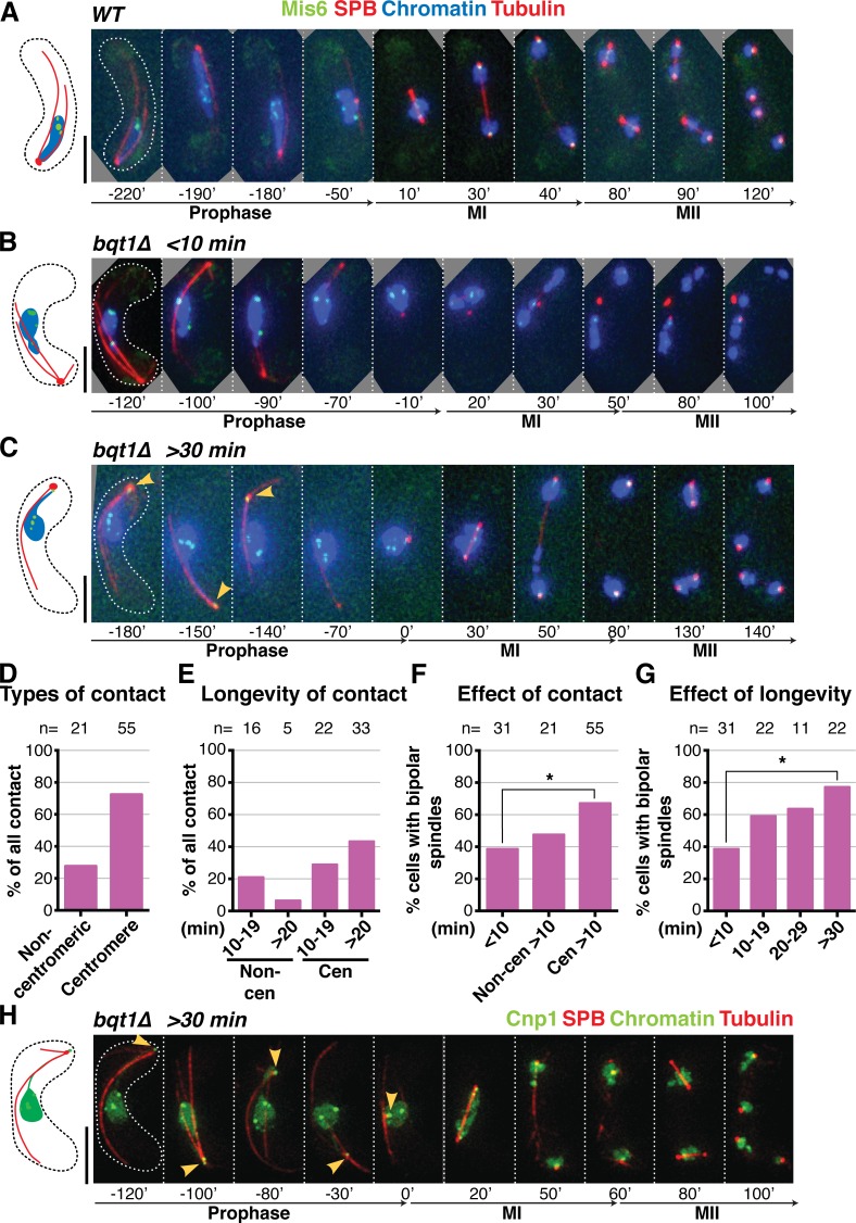
Figure 2.
Centromeres mediate the chromatin–SPB contacts that rescue bqt1Δ spindle defects. (A–C and H) Frames from films of meiocytes carrying Hht1-CFP (at a single endogenous locus as in Fig 1; Chromatin), Sid4-mCherry (SPB), ectopically expressed mCherry-Atb2 (Tubulin), and endogenously tagged Mis6-GFP (A–G) or Cnp1-GFP (ectopically expressed under control of endogenous promoter; H). Bars, 5 µm. (A) In WT cells, centromeres do not localize to the SPB during meiotic prophase. (B) bqt1Δ cell showing <10-min centromere–SPB contact during prophase followed by failed spindle formation. (C and H) A centromere–SPB contact lasting >30 min (indicated by yellow arrowheads) is followed by bipolar spindle formation. (D) Levels of centromeric versus noncentromeric chromatin–SPB contact during bqt1Δ meiotic prophase. (E) Longevity of centromeric versus noncentromeric contacts. (F) Levels of bqt1Δ bipolar MI spindle formation seen in cells with the specified types of chromatin–SPB contact. The percentage of bipolar spindle formation seen in noncentromeric >10-min contact (Non-cen >10 min) is likely an overestimate caused by the faintness of Mis6-GFP signals. (G) Proper spindle formation is quantified as a function of centromere–SPB contact duration. n is the number of cells scored in 14 independent experiments. All cells scored for this figure are more extensively analyzed in Fig. S1 (E and F). *, 0.01 < P < 0.05.

