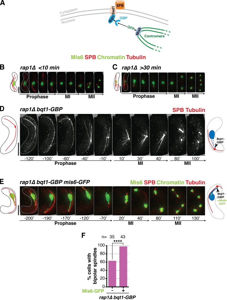
Figure 3.
Maintaining centromeres at the SPB for the entirety of prophase ensures spindle formation in the absence of the bouquet. (A) Schematic of the GBP-GFP system used to force centromere–SPB interactions. (B–E) Frames of films of meiocytes harboring the specified tags: SPB, Mis6, chromatin, and tubulin tagged as in Fig 2; Bqt1 is endogenously fused at its C terminus with GBP. Schematics on the right of each series show the expected prophase phenotype (nuclear membrane outlined with a dashed blue line). (B) In rap1Δ cells lacking centromere–SPB contacts, Bqt1 localizes to the nonchromatin-associated SPB and spindle formation is defective. (C) A rap1Δ meiocyte with >30-min centromere–SPB contact forms proper bipolar spindles. (D) Introduction of Bqt1-GBP in rap1Δ cells lacking a GFP tag on Mis6 fails to rescue abnormal spindle formation. (E) Introduction of Mis6-GFP in the Bqt1-GBP setting confers association of the SPB with a centromere, resulting in proper bipolar spindles. Bars, 5 µm. (F) Levels of bipolar MI spindle formation in rap1Δ bqt1-GBP background with and without Mis6-GFP. n is the total number of cells scored from more than two independent experiments. ****, P < 0.0001.

