Figure 6.
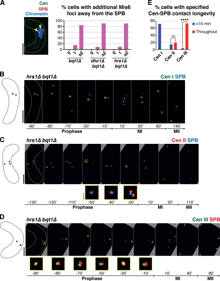
Prophase centromere–SPB contacts in a bqt1Δ setting show a preference for cenIII. (A) In cells with centromere–SPB contacts, additional centromeric foci are observed away from the SPB. Arrowheads mark Mis6-GFP dots far from the SPB during dhc1Δ bqt1Δ prophase. Quantitation is shown to the right. More than 100 cells were scored in five independent experiments. (B–D) Series of frames of films of hrs1Δ bqt1Δ meiosis. Numbering as in Fig 1; the SPB is viewed via endogenously tagged Sad1-CFP. Bars: (black) 5 µm; (gray) 1 µm. (B) The centromere of chromosome I is visualized via a LacI-GFP–bound lys1+-lacO array. (C) The centromere of chromosome II is visualized via a TetR-bound cnt2-tetO array. (D) The centromere of chromosome III was followed via a LacI-bound ade6-lacO array. In these cells, Sad1 is endogenously tagged with mRFP. (E) Collated centromere–SPB interactions. SPB interactions with cenIII are more frequent and longer lasting than those with cenI or cenII; moreover, cenII–SPB interactions are more frequent and longer than cenI–SPB interactions. More than five independent experiments were performed. All cells scored for this figure are more extensively analyzed in Fig. S1 (H–J). ****, P < 0.0001.

