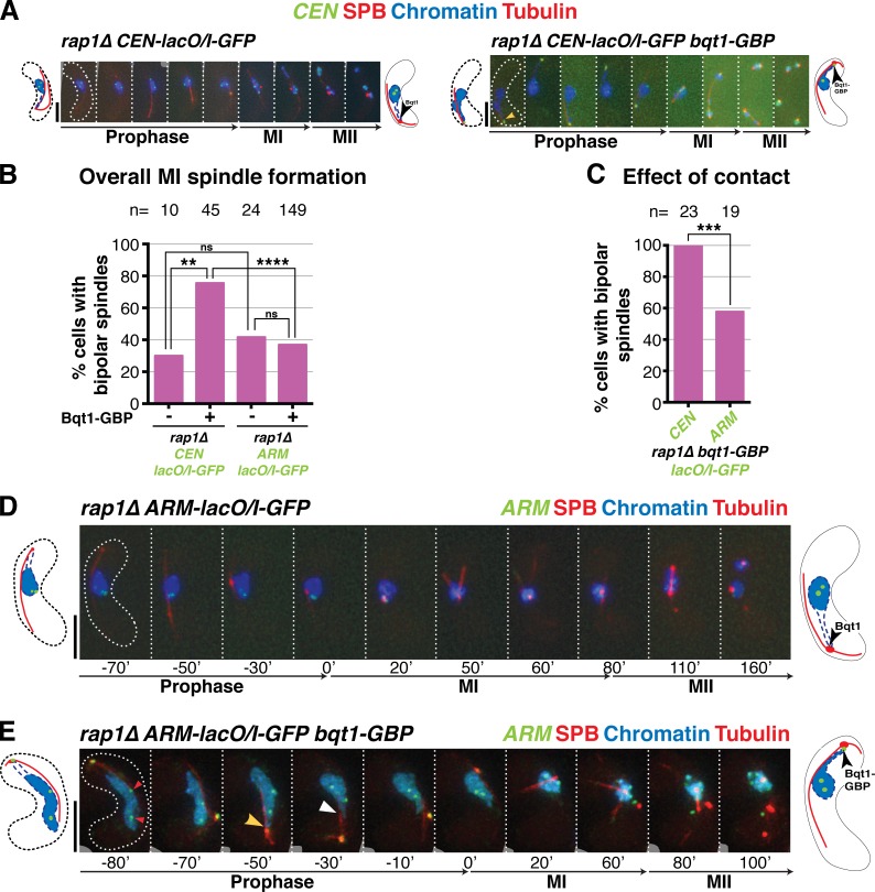
Figure 7.
CEN-proximal regions show greater affinity for the SPB, and greater ability to rescue spindle formation, than ARM-proximal regions. (A, D, and E) Series of frames of films of meiocytes harboring the tags detailed in Fig. S4; numbering as in Fig. 1. Bars, 5 µm. (A) rap1Δ meiotic spindle defect is rescued by forcing long-lived interaction between CEN-proximal region and SPB during meiotic prophase. (B) Quantitation of overall bipolar meiotic spindle formation in specified backgrounds. (C) Quantitation of effect of specified contact on bipolar spindle formation, scoring only those cells with contact >50 min. For meiocytes harboring cut3+-lacO/I-GFP, cells with one GFP focus (n = 4) or two foci in nucleoplasm were scored if and only if a clear GFP focus was present at the SPB. n is the total number of cells scored; data were subject to Fisher’s exact test: ****, P < 0.0001; ***, 0.0001 < P < 0.001; **, 0.001 < P < 0.01. (D) As expected, rap1Δ cells with no chromatin contact have defective meiotic spindles. (E) Example of a rap1Δ cut3+-lacO/I-GFP bqt1-GBP zygote. One GFP focus is seen at the SPB and two are seen within the bulk of the nucleus (left, red arrowheads), indicating incomplete recruitment of cut3 locus to the SPB. Yellow arrowhead indicates clear chromatin–SPB contacts; white arrowhead indicates occasions of less clear contact, in which the nucleus appears to poke out in the direction of the SPB but chromatin markers are not clearly visible.

