Figure 5.
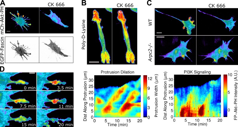
Arp2/3-driven protrusion amplifies adhesion-based signaling. (A) TIRF images of a NIH 3T3 cell coexpressing mCherry-AktPH and EGFP-fascin before and after inhibition of Arp2/3 complex by 50 µM CK666 (representative of 15 cells in six independent experiments). (B) CK666 treatment (100 µM) does not ablate localized PI3K signaling in FP-AktPH–expressing NIH 3T3 cells plated on poly-lysine (representative of 42 cells). (C) FP-AktPH–transfected fibroblasts with conditional knockout of Arpc2 (p34) were either uninduced (wild type [WT]) or induced with tamoxifen (Arpc2−/−). TIRF images acquired before and after CK666 treatment are shown (see also Video 8; representative of 13 cells each). (D, left) Protrusion dilation waves as shown by pseudocolored mCherry-Akt-PH (see also Video 9), representative of 11 such sequences viewed in six cells. (right) The protrusion width and FP-AktPH intensity are plotted for the same sequence on kymograph-like plots. A.U., arbitrary unit. Bars: (A–C) 20 µm; (D, left) 10 µm.

