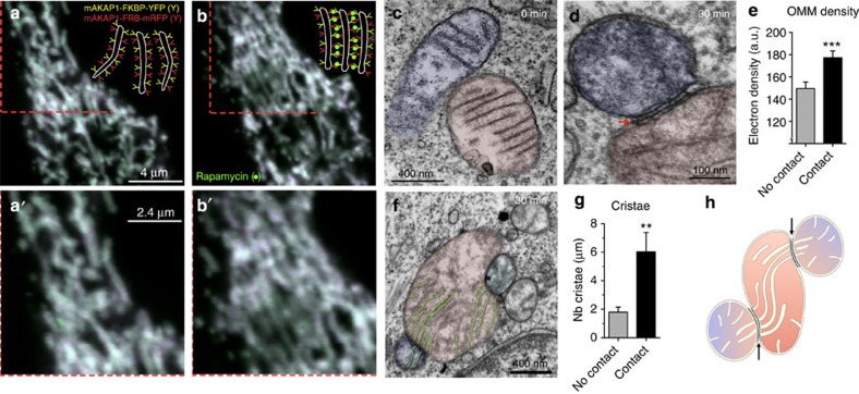
Figure 5. Mito–mito linker induces IMJs and coordination of cristae.
(a,a′) Confocal imaging of RBL-2H3 cells transfected with mitochondria-targeted inducible linkers at 0 min and (b,b′) 30 min post-induction with rapamycin. (c,d) Electron micrographs from (c) non-induced and (d) 30 min post-induction of mitochondrial linkage, (e) showing increased electron density selectively at site of contacts (means±s.e.m., paired T-test, n=20 per group). (f,g) Quantification of cristae abundance at linker-induced contact sites compared with no contact mitochondrial surfaces (means±s.e.m., paired T-test, n=14 per group). (h) Theoretical model whereby cristae organization and density are regulated at IMJs, possibly enabling the equilibration of the membrane potential across physically tethered organelles. **P<0.01, ***P<0.001.

