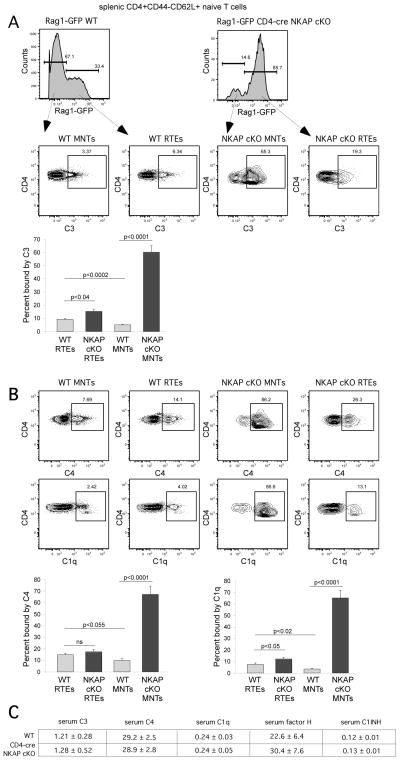
FIGURE 2.
Increased complement C3 and C4 deposition, and binding of complement C1q, is found in peripheral naïve T cell population of CD4-cre NKAP cKO mice. Splenocytes from Rag1-GFP WT and Rag1-GFP CD4-cre NKAP cKO mice were incubated in the GVB++ buffer prior to staining with anti-C3 or anti-C4 (A), or anti-C1q antibodies (B). Splenic CD4+CD44−CD62L+Rag1-GFP+ cells were defined as RTEs while CD4+CD44−CD62L+Rag1-GFP− cells were defined as mature naïve T cells (MNTs). Data shown are representative of at least 4 mice per group from 7 independent experiments. Quantitation from at least 7 Rag1-GFP WT and 7 Rag1-GFP CD4-cre NKAP cKO mice from at least 5 independent experiments is also shown. Error bars indicate SEM. Data were analyzed for significance by an unpaired Student’s t test. NS, not significant. (C) Serum concentrations of C3, C4, C1q, factor H and C1INH in 8 WT and 7 CD4-cre NKAP cKO mice were determined by ELISAs. The average concentration in μg/ml and the SEM for each is shown. The concentrations of these proteins in WT and CD4-cre NKAP cKO mice were very similar.

