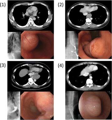
Figure 4.

Findings of contrast-enhanced computed tomography, barium-swallow esophagogram and endoscopy in four cases. (1) Gastrointestinal stromal tumor located on the left side of the middle third of the thoracic esophagus. (2) Tumor located on the lower third of the esophagus was histopathologically diagnosed as leiomyoma. (3) Large tumor located on the lower third of the esophagus was histopathologically diagnosed as leiomyoma. (4) Tumor located on the lower third of the esophagus was histopathologically diagnosed as leiomyoma.
