Figure 2.

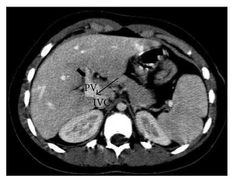
Axial contrast-enhanced CT images. The arrow shows the shunt between the portal vein (PV) and the inferior cava vein (ICV); at the hepatic hilum, the PV appears enlarged with only one intrahepatic portal branch (arrowhead).

Axial contrast-enhanced CT images. The arrow shows the shunt between the portal vein (PV) and the inferior cava vein (ICV); at the hepatic hilum, the PV appears enlarged with only one intrahepatic portal branch (arrowhead).