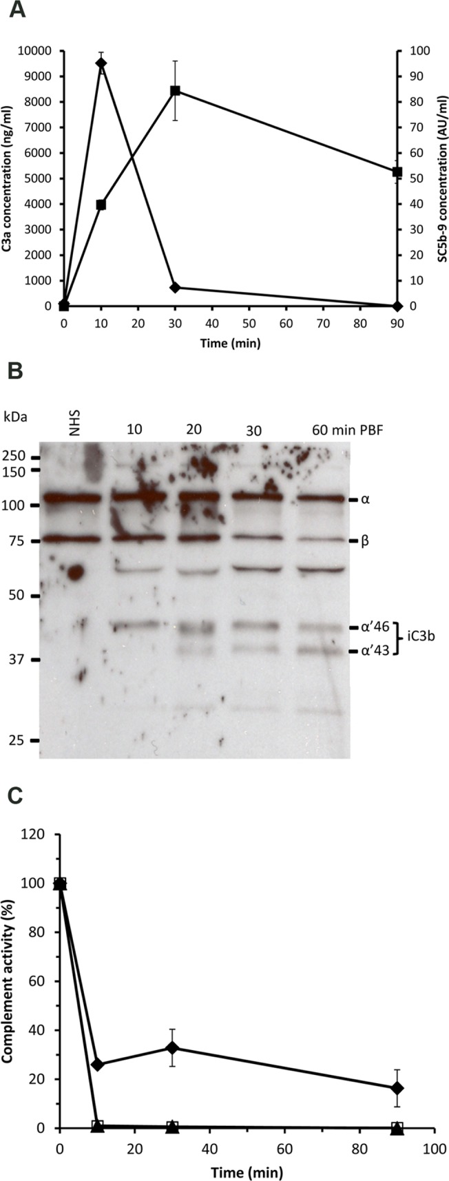
Fig 1. Complement activation in the A. stephensi mosquito midgut after feeding on blood from the arm of a human volunteer.

(A) The concentrations of the complement activation products C3a (♦) and SC5b-9 (■) were measured in sera collected from mosquito midguts at 10, 30 and 90 min PBF on a human volunteer arm. C3a and C5b-9 were also measured in a serum sample (zero time) of the volunteer. C3a level peaked at 10 min PBF and then dropped sharply at 30 min PBF, whereas the C5b-9 level peak was reached later at 30 min PBF. (B) Conversion of C3 into iC3b as a sign of regulated complement activation was assessed by Western blot analysis. Serum samples taken before blood feeding and collected from mosquito midgut at 10, 20, 30 and 90 min PBF showed decreasing intensities of C3 α-chain and increasing intensities of α’ 46 and α’43 kDa chains (iC3b fragments) of over time. (C) Residual complement activity in serum samples collected from mosquito midguts at 10, 30 and 90 min PBF. Both the alternative (AP; ▲) and the lectin pathways (LP; □) of complement activation appeared to be completely inhibited or consumed at 10 min PBF, whereas about 20% of the classical pathway (CP; ♦) activity was still detectable at 90 min PBF.
