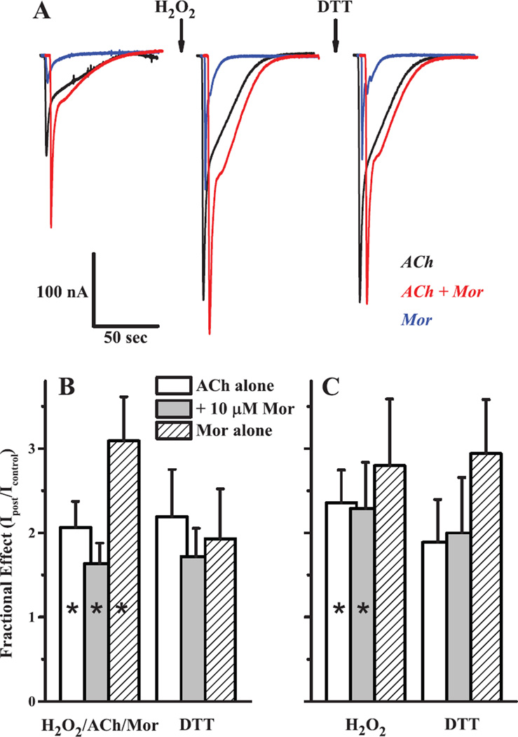
Figure 7.
Oxidation of the α3Q37Cβ2A127C receptor increases activation. The effects of sequential oxidation and reduction treatments on α3S125Cβ2Q39C receptors are shown as representative sample traces (A) and as collated data for currents after treatment normalized to the original (naïve) controls using two oxidation treatments of (B) 4.4 mM H2O2 + 100 µM ACh + 10 µM Mor and (C) 4.4 mM H2O2 alone. In panel A, currents were evoked by 1 mM ACh alone (black), 1 mM ACh + 10 µM Mor (red) or 10 µM Mor alone (blue) with the oxidation treatment (first arrow) as 5 min 4.4 mM H2O2 and reduction (second arrow) with 5 min of 40 µM DTT. For panels B and C, collated data of the type shown in panel A for currents evoked by ACh alone (open bars), ACh + Mor (gray bars) and Mor alone (hashed bars), at the concentrations given above, are plotted as bar graphs; reduction treatment was with 40 µM DTT in both cases. Bars represent means (+ SEM). For experiments of n = 7 (H2O2/ACh/Mor; all three challenges in B), n = 5 (DTT in B) and n = 4 (both treatments; all three challenges in C), all changes upon oxidation were significant at p < 0.05 (range 2.5×10⁻⁵−0.035), except C. Mor alone (p = 0.132), and are denoted with an asterisk. DTT did not produce significant changes in any of the three currents for either oxidation treatment (p = 0.096–0.457).

