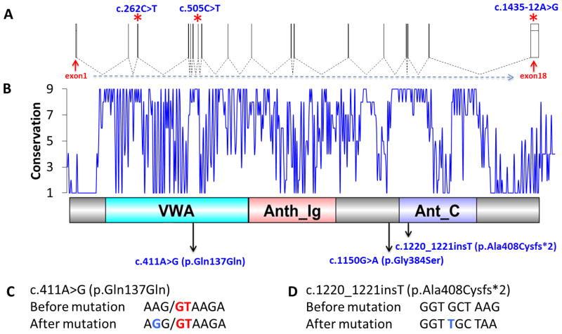
FIG. 3.
Gene structure and diagram of the functional domains of ANTXR1 and localization of the identified mutations. A: Demonstration of ANTXR1 exons. Asterisks indicate localization of previously described mutations in three different ethnicities (c.262C>T = Egyptian, c.505C>T = Czech, c.1435-12A>G = Sri Lankan). B: Conservation pattern and protein domains of ANTXR1 (564 aa). Conservation varies between 1 (variable) and 9 (conserved). Arrows indicate localization of the amino acid changes described in our study in three Turkish families. C: c.411A>G (p.Gln137Gln) mutation. G (blue) and GT (red) are substitution and canonical splicing donor site. D: c.1220_1221insT (p.Ala408Cysfs*2) mutation. Inserted T is highlighted by blue.

