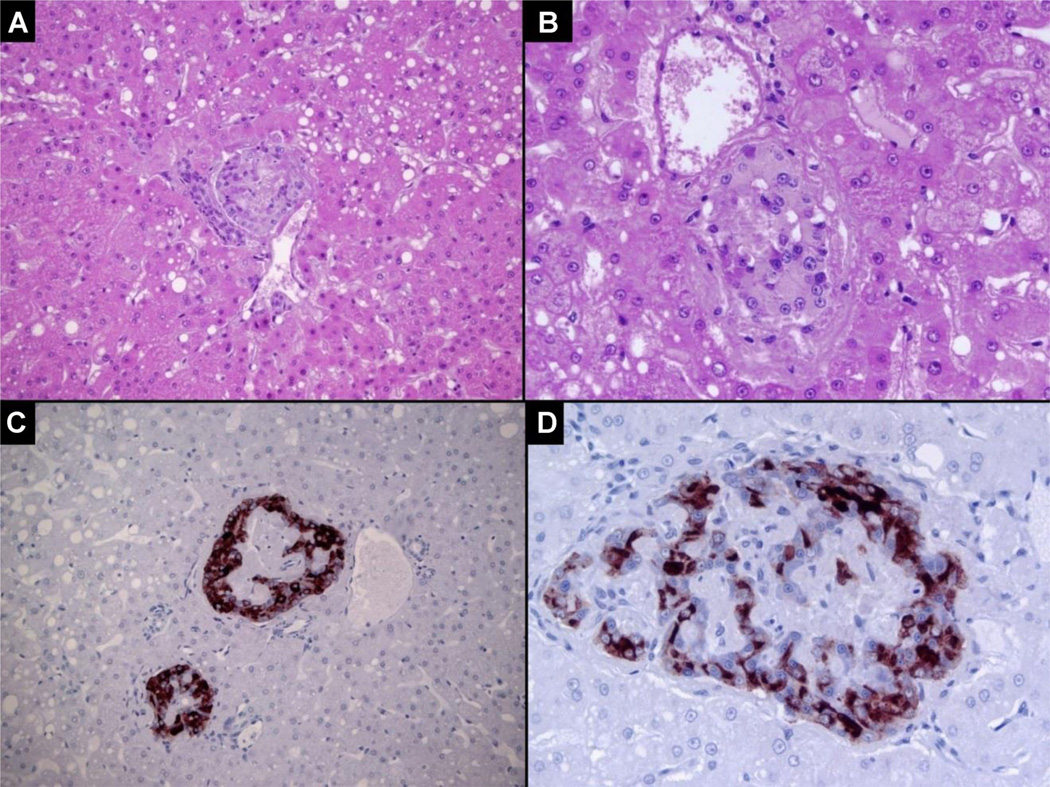
Figure 2. Histological examinations of islets from an alpha-1-antitrypsin-treated autograft recipient 468 days posttransplantation. Serial liver samples were fixed prior to sectioning and staining.
The following was observed: (A) Islets present in a small portal vein with mild fibrin deposition. Micro-steatosis is evident. Hematoxylin and eosin (H&E), 200×; (B) Islet in a small portal vein with minimal fibrin deposition and hepatic micro-steatosis. H&E, 400×; (C) 200× and (D) 400× islets in small portal venules with strong insulin staining detected by immunohistochemistry. The sections were counterstained with hematoxylin.

