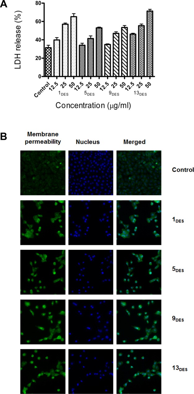
Fig 5. DESs treatment increase LDH release and membrane permeability.
(A) LDH% release by MCF-7 cells treated with various dosages of DESs. After 48 h treatment, the released LDH were quantified by a coupled enzymatic reaction, and measured using a fluorescence plate reader. (B) MCF-7 cells treated with LC50 dosages of DESs for 24 h. Cells were fixed and stained with membrane permeability dye (green) and nucleus was stained with Hoechst 333258 dye (blue).

