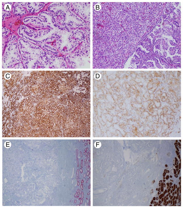
Fig. 1.
Histologic and immunohistochemical features of CCPRCC. A, Tumor cells showed typical nuclear arrangement away from the basement membrane. B, Small tubules and acini imparting a solid appearance admixed with delicate papillary structures. C, CK7 showed strong and diffuse positivity. D, CAIX showed the typical basal staining lacking the luminal aspect. E, AMACR resulted negative in all cases; normal distal tubules were positive. F, CD10 was negative or weakly positive. A, Original magnification HE, ×20; B, HE, ×10; C to F, ×10.

