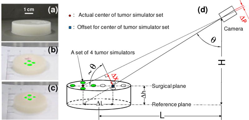
Figure 4.

Correction of the co-registration error between the fluorescence image of the tumor margin and the surgical scene image. (a) Photo of agar-agar gel phantom with four embedded tumor simulators. (b) Without correction, the co-registration error between the fluorescence image (green) and surgical scene image is significant. (c) With height correction, the center of the projected tumor simulator set matches the actual position.
