
Figure 8.
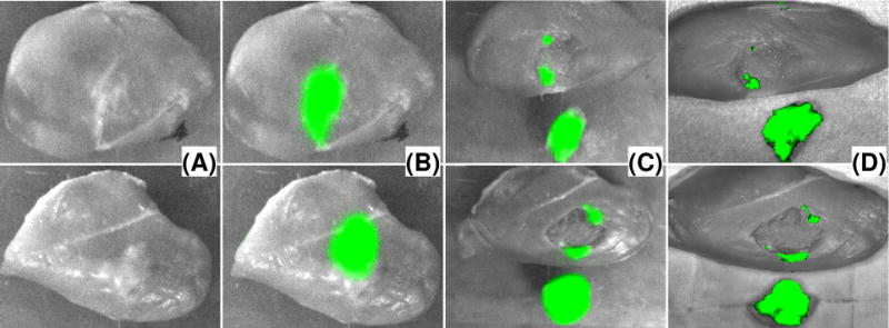
Image-guided tumor resection surgery on an ex vivo tumor-simulating model. Top row are images for Google glass guided surgery. Bottom row are images for HMD guided surgery. (A) Photographic images of the surgical field without fluorescence filter. (B) Fluorescence images of the surgical margins before resection. (C) Fluorescence images of the surgical margins and the resected tissue samples after resection. (D) IVIS image of the resected tissue sample and the surgical cavity.