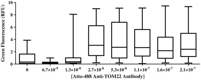
Fig. 4.
Distribution of individual fluorescence intensities of dual-labeled events. The box plot presents the 25th, 50th, and 75th percentile and whiskers represent the minimum and maximum values of green fluorescence (RFU) of dual-labeled events. At concentrations of 2.7 × 10⁻⁸ to 2.1 × 10⁻⁷ M antibody, there is a significant increase in RFU of green fluorescence in dual-labeled events (P<0.0001, α=0.05)

