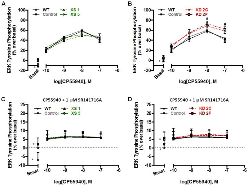
Figure 5. Efficacy of CP55940-stimulated phosphoERK1/2 is enhanced by depletion of CRIP1a.
N18TG2 WT, empty vector (Control), CRIP1a XS (A,C), and CRIP1a KD (B,D) cells were serum-starved for 16 h, pretreated for 2 h with 1 μM THL, and treated with varying concentrations of CP55940 (A,B), or CP55940 in the presence of 1 μM SR141716A (C,D) for 5 min. In-cell-Western analysis of phosphoERK1/2 was quantified as the ratio of phosphoERK1/2:total ERK, and then phosphoERK values were presented as a percent change relative to basal for each clone. Data are presented as the mean ± S.E.M. calculated from three independent experiments performed in duplicate. #p<0.05 indicates that data points significantly differ from WT, using a two-way ANOVA and Bonferroni post-hoc test. *A significant difference from basal was observed for CRIP1a XS and KD clones, as indicated in Fig. 1A, B, C and D, respectively.

