
Figure 6.
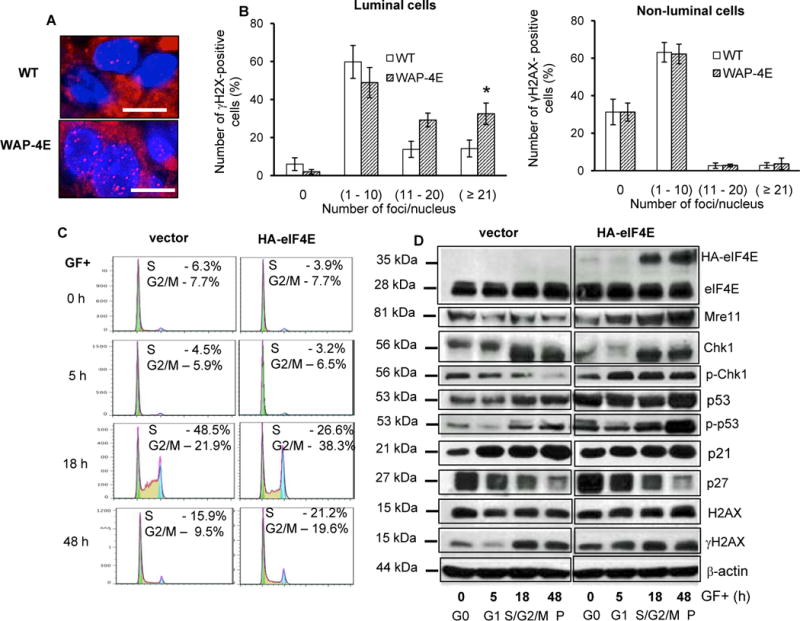
Overexpressed eIF4E accelerates cell cycle progression and stimulates a replication stress response. A and B – analysis in vivo: A, representative images of mammary epithelial cells from wild-type (WT, n=3)) and transgenic (WAP-4E, n=4) mammary glands (gestation 6). Sections were immunostained for γH2AX (red) and DAPI (blue). Bars =10 μm. (B), quantification of γH2AX expression in luminal and non-luminal cells (mean ± SE, n=4), *p ≤ 0.001. C and D – analysis ex vivo: C, representative flow cytometric histograms showing cell-cycle progression of HMEC/hTERT cells harboring HA-eIF4E or an empty vector. Cells were growth factor restricted for 48h, stimulated to cycle with growth factors, and DNA content was analyzed at the indicated times post-growth factor stimulation. D, whole-cell lysates were obtained at the indicated times post-growth factor stimulation of cultured cells and immunoblotted for cell cycle checkpoint regulators (p27, p53, p-p53Ser15 and p21), ATR targets (Chk1, H2AX, γH2AXSer139) and Mre11. β-actin was used as a loading control. P- asynchronously proliferating cells. Shown are represented histograms (C) and blots (D) from 3 independent experiments