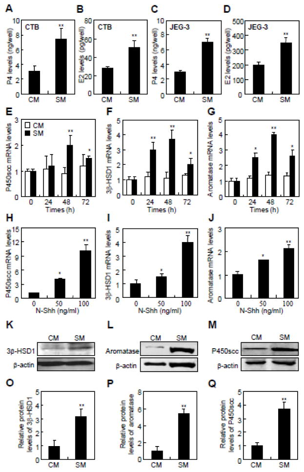
Fig. 1.
Hh signaling stimulates cholesterol conversion and steroidogenic enzymes expression in primary cytotrophoblasts and trophoblast-like JEG-3 cells. (A–D) P4 and E2 levels in the culture media of cytotrophoblasts (CTBs) and JEG-3 cells containing 25-hydroxycholesterol and testosterone, respectively, after 48 hrs of either control medium (CM) or Shh conditional medium (SM) treatment. (E–J) Determination of mRNA levels of P450scc, 3β-HSD1, and aromatase by quantitative RT-PCR in JEG-3 cells following CM, SM or Shh recombinant protein (N-Shh) treatments. (K–M) Measurements of the expression of P450scc, 3β-HSD1, and aromatase by western blots in JEG-3 cells following the same treatments. (O–Q) Quantification via densitometry (n=3) and statistical analysis of bands of K–M, respectively. The data shown represent average fold of Shh-treatment groups compared with the control groups. RNA and protein abundance normalized to β-actin, respectively. **p<0.01, *p<0.05; n=6, error bar, SD.

