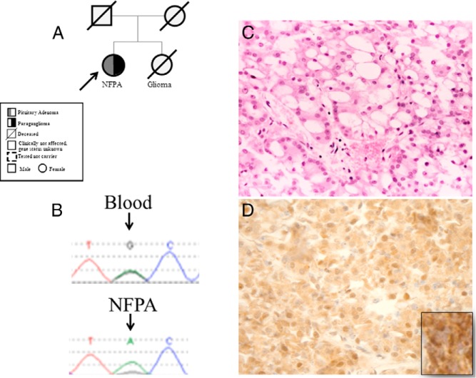
Figure 2.
Pedigree (A) and LOH (B) at the SDHB locus in the pituitary adenoma of patient 4; the microsatellite upstream of the mutation has also shown to be lost. C, H&E-stained section (×20) of this adenoma shows prominent vacuolar changes in most neoplastic cells; the cytoplasm otherwise appears weakly eosinophilic. D, SDHB staining suggesting lack of strong granular staining of the pituitary adenoma of the proband (immunoperoxidase, ×20) (inset: positive SDHB staining as positive control in a paraganglioma).

