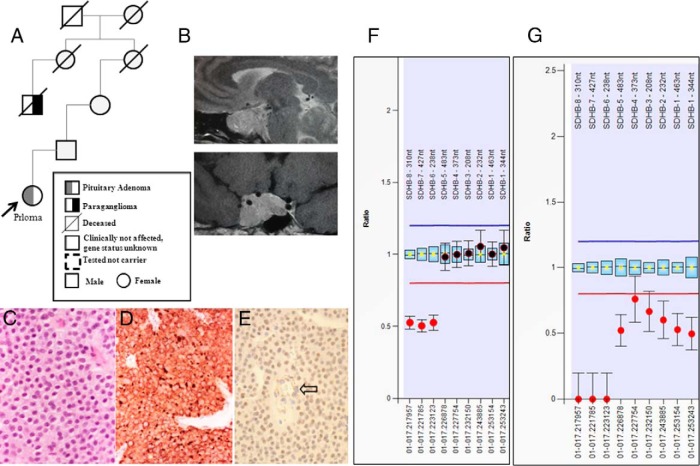
Figure 3.
Pedigree (A) and sagittal and coronal magnetic resonance images of the pituitary adenoma (B) are shown. C, H&E-stained section (×20) shows that the tumor of patient 5 contains multiple vacuoles. D, The immunoreaction with the anti-113-1 antibody (immunoperoxidase, ×20) highlights the mitochondria content. E, SDHB immunostaining shows loss of expression in neoplastic cells, whereas endothelial cells (arrow) retain the expression (immunoperoxidase, ×20). Loss of the SDHB gene in germline and pituitary tumor tissue in patient 5. F, Germline DNA shows a deletion affecting MLPA SDHB probes 6–8 in DNA derived from leukocytes. G, In pituitary adenoma tissue, a complete loss of genetic material at the SDHB probes 6–8 area and heterozygous loss of SDHB probes 1–5.

