Figure 2.
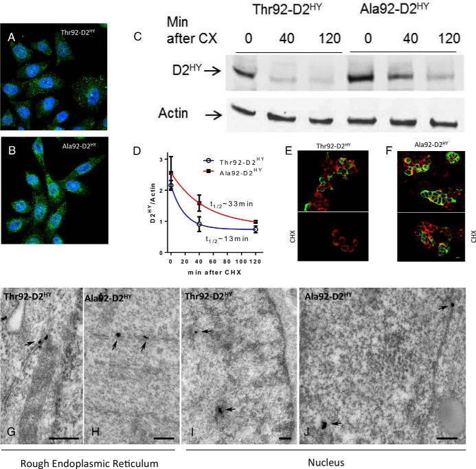
Thr92–D2HY and Ala92–D2HY are both found in the nucleus but Ala92–D2HY has a longer half-life. A, B, Immunofluorescence of HEK-293 cells stably expressing D2HY proteins; D2 was stained with αYFP (green) and is found throughout the ER; nuclei were stained with DAPI (blue); (C) the indicated cells were incubated for 40–120 minutes with 100 uM CHX and subsequently harvested and processed for western analysis using αYFP and αActin; (D) quantification of the bands shown in C; data were analyzed by nonlinear regression assuming that D2 decays exponentially after addition of CHX; all data are the mean ± SEM of three entries per data-point; (E–F) immunofluorescent detection of plasma membrane by the Na+/K+-ATPase (red) and αYFP of D2HY fusion proteins (green) in the same cells as A-B before (above) and after (below) treatment with CHX for 120 minutes; (G) EM of HEK-293 cells stably expressing D2HY proteins as in A-B; silver grains denoting D2HY can be visualized in the rough endoplasmic reticulum (arrows) in both Thr92–D2HY and (H) Ala92–D2HY-expressing cells; (I) the nucleus also contains Thr92–D2HY and (J) Ala92–D2HY proteins; scale bars = 0.25 um.

