Figure 3.
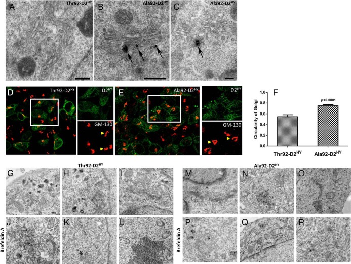
Ala92–D2HY, but not Thr92–D2HY, can be found in the Golgi. A, Electron microscopy of Thr92–D2HY expressing cells where a typical Golgi apparatus is devoid of D2 protein; (B, C) same as A except that Ala92–D2HY expressing cells were studied; in this case, silver grains denoting the presence of Ala92–D2HY protein are observed associate to the Golgi apparatus (arrow); scale bars = 0.25 um; (D) Immunofluorescence staining of Thr92–D2HY and (E) Ala92–D2HY (green) expressing cells with cis-Golgi marker GM-130 (red). White box is enlarged in green (D2) and red (GM-130) channels. Yellow arrows show Thr92–D2HY and Ala92–D2HY specific cis-Golgi staining features, where Ala92–D2HY Golgi demonstrates a circular morphology compared to the ribbon configuration in the Thr92–D2HY-expressing cells; (F) The circularity index of the cis-Golgi complex in individual D2HY-expressing cells was measured in ImageJ where the cis-Golgi structure in Ala92–D2HY cells had higher circularity values than from Thr92–D2HY cells; (G-I, M-O) Untreated cells display the typical appearance of Golgi-complex independently from the type of D2 expressed in the cells. The cisternae of the Golgi complex were organized in parallel, slightly curved and surrounded by small Golgi vesicles; (J-L) In cells expressing the Thr92–D2HY, BFA treatment (0.5 μg/mL) resulted in a disorganization of the Golgi apparatus with scattered, dilated and short cisternae. However, some organized Golgi can be observed; (P-R) In BFA-treated Ala92D2HY-expressing cells, circular Golgi complexes were present that were otherwise unidentified in Thr92–D2HY expressing cells.

