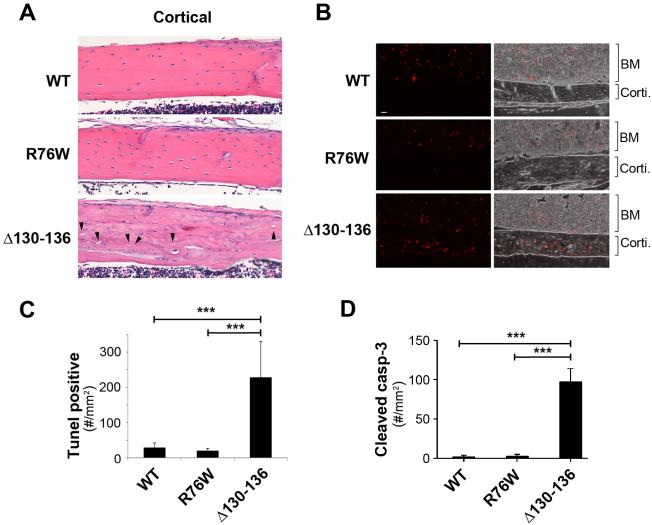
Figure 5. Apoptotic osteocytes and empty lacunae are increased in trabecular and cortical tibial bone of Δ130-136 mice.
(A) H&E staining of paraffin sections of 4-month-old tibial bones showed increased numbers of empty lacunae relative to total lacunae in tibial cortical bone in four-month-old Δ130-136 mice, but minimal differences in WT as compared to R76W mice. The solid arrowheads point to the empty lacunae. (B) Paraffin sections of 4-month-old tibial bones were TUNEL stained showing fluorescence TUNEL images (left panels) and overlay corresponding fluorescence and phase images (right panels). A significant increase of TUNEL signals was detected in cortical bone area (corti) of Δ130-136 mice, but not much in that of WT and R76W mice. Comparable TUNEL signals were detected in bone marrow (BM) of WT and two transgenic lines. (C) The TUNEL signals per mm2 of cortical bone area were quantified. (D) Paraffin sections of 4-month-old tibial bones were immunolabeled with anti-cleaved caspase-3 antibody and the numbers of cleaved caspase-3-positive osteocytes per mm2 of cortical bone area was quantified. Data shown are mean ± SD. ***, P < 0.001. n = 3.

