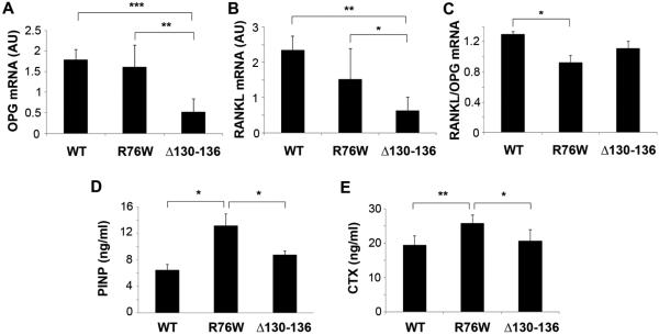
Figure 8. Changes of markers of bone remodeling in transgenic models.
(A-C) Total RNA were extracted from cortical bones of WT and two transgenic mouse models, and were used for RT-PCR analysis probed with primers for OPG and RANKL. mRNA expression of OPG (A) and RANKL (B) was significantly decreased in Δ130-136 compared to WT and R76W mice, while RANK/OPG ratio (C) was reduced in R76W mice compared to WT. (D and E) Sera were collected from WT and two transgenic mouse models, and the concentration of PINP (D) and CTX (E) was determined by ELISA assay. The contents of both PINP and CTX were significantly increased in R76W mice as compared to WT and Δ130-136 mice. Data shown are mean ± SD. *, P < 0.05; **, P < 0.01; ***, P < 0.001. n = 6.

