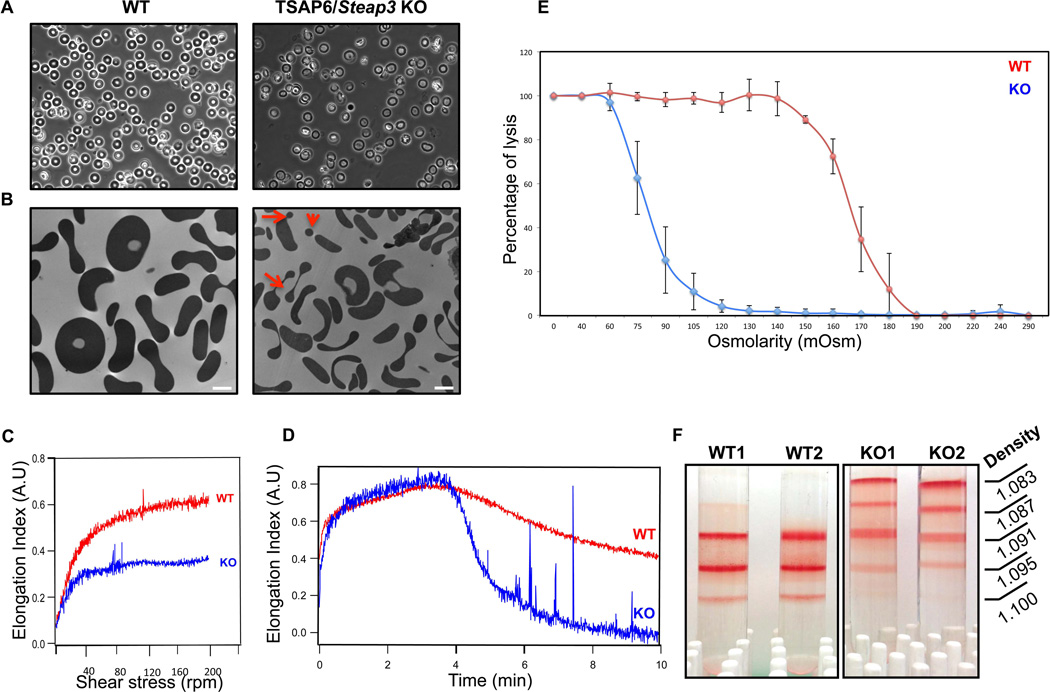
Figure 1. Phenotype of red cells from TSAP6/Steap3 KO mice.
(A) Phase contrast microscopy of peripheral blood from wild type (left panel) or TSAP6/Steap3 knockout mice (right panel) shows heterogeneity in the shape of the red blood cells. (B) This is confirmed by transmission electron microscopy, where fragmentation of erythrocytes is apparent (red arrows). In addition, cells are smaller. Bar represents 2 µm. (C–D) Membrane deformability assessments by ektacytometry in 3% PVP (C) or 40% dextran (D) show reduced deformability for TSAP6/Steap3 KO red blood cells. (E) Osmotic fragility is also reduced in knockout animals. Data presented X ± Standard deviation, n=4 in each group. All animals were 4 months of age. (F) Density gradients performed on red cells show a different density distribution in the knockout samples.

