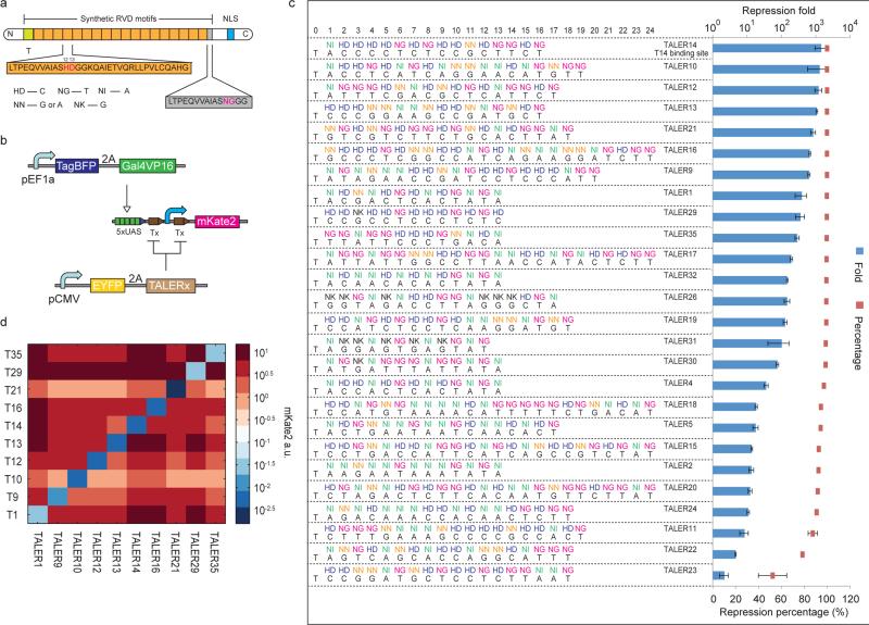
Figure 1. Design and construction of TALE repressors for mammalian cells.
(a) Schematic representation of TALE repressors with repeat di-variable residue (RVD) domains. The repeat domains are shown in orange blocks with RVDs in magenta and the binding specificity of RVDs is shown below. The last domain is a 15 amino acid half repeat domain depicted in a grey box. The green block represents the requirement of a leading repeat that binds T. The blue block represents a nucleus localization signal (NLS). (b) Design of a fluorescent reporter assay for measuring TALER repression fold and orthogonality. Tx: binding site for TALER x. Blue arrow represents a minimal CMV promoter. Lines with arrows indicate up regulation and lines with bars indicate down regulation. (c) Functional assay of TALERs in HEK293 cells. Selected RVD domains and binding sequences are shown on the left. Blue bars represent repression fold and red boxes represent repression percentage. Each bar/box shows mean ± SD from three independent flow cytometry experiments. (d) TALER orthogonality matrix. Each box in matrix represents fluorescent reporter expression measured in co-transfection of indicated TALER protein and promoter.

